Symptoms & diagnostics
Cytology or biopsy: how tumour tests differ
Cytology and histopathology answer different questions. What each test shows, how long results take, why a biopsy before surgery can matter, and what to do if the answer is unclear.
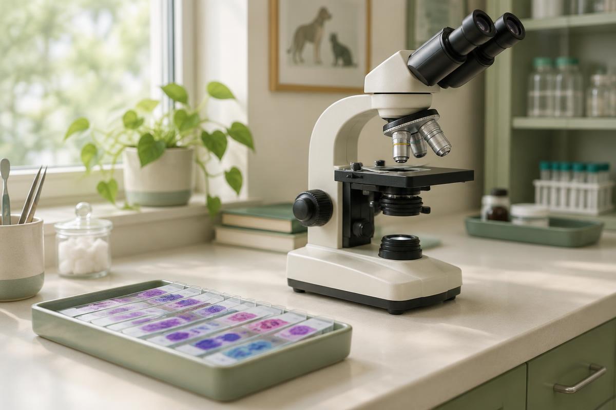
To find out what a growth is, its cells or tissue need to be examined under a microscope. There are two main routes. Cytology looks at individual cells collected with a fine needle — it's quick and minimally invasive. Biopsy with histopathology looks at a piece of tissue — it takes longer and is more involved, but gives more information. They are often used in sequence: cytology first, then a biopsy if needed.
The short answer
- Cytology often gives an answer for benign lumps and for some tumours, such as lymphoma and mast cell tumours.
- Histopathology identifies the exact type of most tumours, their grade, and — after removal — the surgical margins.
- An inconclusive result isn't a failure; it points to the next step.
- Knowing the diagnosis before surgery helps the surgeon plan how much to remove.
What cytology is
In a fine-needle aspirate (FNA), the vet inserts a thin needle into the lump and collects a small number of cells, which are spread on a slide and stained. Anaesthesia is usually not needed. Your vet may read the slides in the clinic or send them to a clinical pathologist — the latter is recommended when there's doubt, or when the result will shape treatment. Results usually take zero to two days.
Cytology shows whether a lump is inflammation or a tumour and, if it's a tumour, which broad group it belongs to: epithelial, mesenchymal or round cell. Sometimes it can also suggest whether it is benign or malignant.
What biopsy and histopathology are
A biopsy collects a piece of tissue, which is fixed and examined by a veterinary pathologist. Options include:
- Incisional biopsy — part of the mass is sampled, with a special needle (needle-core or "Tru-Cut"), a circular punch or a wedge incision.
- Excisional biopsy — the whole mass is removed and sent for examination.
A biopsy usually needs local anaesthesia, sedation or general anaesthesia, depending on the site and technique. Histopathology results typically take three to seven days.
Side by side
| Cytology | Histopathology | |
|---|---|---|
| How it's taken | Fine needle, usually no anaesthesia | Tissue sample or whole mass, anaesthesia needed |
| Turnaround | Usually 0–2 days | Usually 3–7 days |
| What it shows | Cell type, sometimes benign vs malignant | Exact type of most tumours, grade, invasion |
| Surgical margins | Not assessed | Assessed after removal |
Why a result can be inconclusive
Some tumours, such as soft tissue sarcomas, don't shed many cells onto a needle. A sample may contain mostly blood or fat, and in a large mass the needle may hit an area of inflammation or dead tissue. Your vet may then repeat the aspirate or suggest a biopsy. If cytology and histopathology disagree, histopathology usually carries more weight, but your vet will interpret it in the context of the whole picture.
Additional tests
When standard methods aren't enough, a pathologist or oncologist may suggest immunohistochemistry, special stains, PCR for antigen receptor rearrangement (PARR) or flow cytometry. These are most often used to classify lymphoma or to identify the origin of a poorly differentiated tumour.
Why a diagnosis before surgery matters
For some tumours — mast cell tumours and soft tissue sarcomas, for example — the surgeon plans to remove a margin of healthy tissue around the mass. If such a tumour is removed without that plan, the margins may be too narrow and a second operation may be needed. That's also why the biopsy site is planned so it can be removed together with the tumour later.
Are these procedures safe?
FNA is considered minimally invasive. A small bruise can occur, and a mast cell tumour may become temporarily red or swollen after being handled. For certain locations, such as the bladder, vets choose other sampling methods to reduce risks. Ask your vet about the specific risks in your pet's case.
Questions to ask your vet
- Which test do you suggest first, and why?
- Who will read the sample — you or a laboratory?
- What will we do if the result is inconclusive?
- Do we need a diagnosis before surgery in this case?
- Might extra tests such as immunohistochemistry be needed?
Frequently asked questions
Cytology says "benign" — can I stop worrying?
Often this is genuinely good news. But cytology only sees some of the cells, so your vet may recommend monitoring or further tests if the lump changes.
Can a biopsy spread the tumour?
A properly planned biopsy rarely causes spread, and the value of an accurate diagnosis usually outweighs the risk. Discuss any concerns with your vet.
Can I get a second opinion on the slides?
Yes. Laboratories usually keep slides and tissue blocks, and they can be sent to another pathologist for review.
Important
This article is for general information and does not replace advice from a veterinarian. The right test depends on the individual animal and lump. An online consultation can help you understand reports you've received, but it can't replace an exam or sampling.
Sources and further reading
- AAHA Section 3: Tumor Diagnostics and Staging (opens in a new tab)
- AAHA: 2026 Oncology Guidelines for Dogs and Cats (opens in a new tab)
- ACVS: Mast Cell Tumors (opens in a new tab)
- Merck Veterinary Manual: Cancer Treatment in Animals (opens in a new tab)
- Veterinary Cancer Society: Pet Owner Resources (opens in a new tab)
Updated 20 October 2026
Related articles
Symptoms & diagnostics
Mammary Tumors in Dogs and Cats: Signs, Diagnosis and Treatment
A lump near a nipple cannot be assessed by touch alone. Learn how mammary tumors differ in dogs and cats, which tests may be needed, and how surgery and follow-up care are planned.
8 min read
Symptoms & diagnostics
Tumor biopsy in dogs and cats: what happens and why it matters
A biopsy helps identify a tumor and guide treatment. Learn how tissue is collected, what to expect on the day, how to care for the wound, and what the pathology report can tell you.
7 min read
Symptoms & diagnostics
Cancer symptoms in dogs and cats: what to watch for
Signs of cancer in pets are often vague — weight loss, tiredness, a cough or a limp. Which changes to show your vet, which need urgent care, and how to keep useful notes at home.
4 min read