Symptoms & diagnostics
Tumor biopsy in dogs and cats: what happens and why it matters
A biopsy helps identify a tumor and guide treatment. Learn how tissue is collected, what to expect on the day, how to care for the wound, and what the pathology report can tell you.
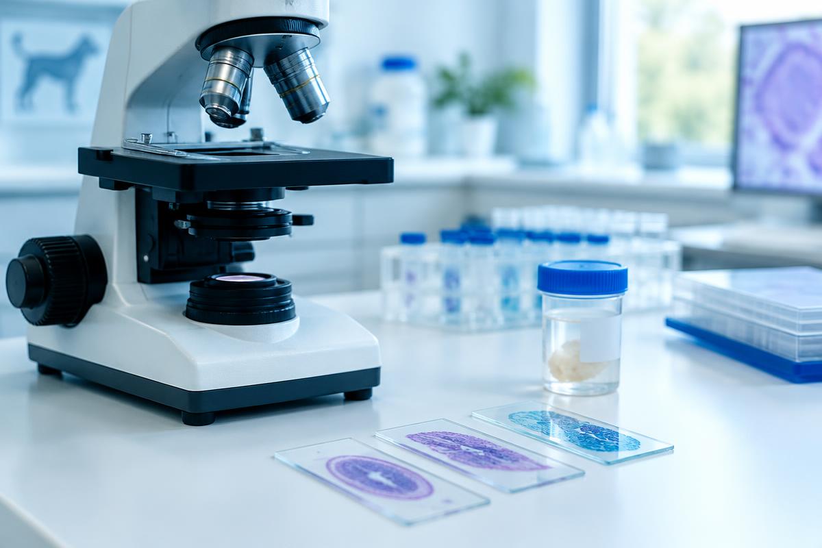
A biopsy collects tissue for examination under a microscope. In dogs and cats, it helps clarify what a lump or abnormal area is before choosing treatment. A recommendation for biopsy does not mean cancer has already been confirmed. The tissue may show inflammation, a benign growth, or malignancy.
The procedure may involve local anesthesia, sedation, or general anesthesia. The choice depends on the location, the amount of tissue needed, and your pet’s health. Planning should also consider any later surgery, not just how to obtain a sample today.
Key points
- A biopsy helps plan treatment, not simply name a tumor.
- The sampling site should fit the plan for possible definitive surgery.
- Preparation and pain relief are tailored to each animal.
- Results often take about a week; additional testing takes longer.
- Protecting the wound and following discharge instructions are important.
Why identify the tumor before treatment?
Lumps that feel similar can need very different approaches: monitoring, surgery with planned margins, medical treatment, or a combination. A photograph, firmness, or growth rate cannot reliably identify the tissue. A new lump on a dog deserves assessment even if it is painless.
Sampling before surgery matters when the diagnosis could change the operation. Suspected soft tissue sarcomas, for example, require thoughtful planning of surrounding tissue removal. For mast cell tumors in dogs, cytology often supplies the initial diagnosis; whether a biopsy is needed and how grade will be assessed are separate decisions. Not every pet needs every test.
Cytology and histology (biopsy): comparison
| Question | Cytology | Histology of biopsy tissue |
|---|---|---|
| Collection | Cells obtained with a fine needle or a smear | A core, tissue fragment, or entire mass |
| Anesthesia | Often unnecessary, depending on site and patient | Usually local anesthesia, sedation, or general anesthesia |
| Results | Often faster; laboratory schedules vary | Commonly about a week, longer for complex cases |
| Information | Cell type and signs of inflammation or cancer | Tumor type, sometimes grade and invasion; margins in suitable specimens |
| Limitations | Does not show tissue architecture or surgical margins | Small samples may miss important areas; not every measure applies |
For a fuller discussion of choosing between these tests, see cytology versus biopsy. The focus here is the biopsy procedure itself.
Types of biopsy
Incisional biopsy: sampling part of the mass
A core needle, sometimes called Tru-Cut, removes a narrow cylinder of tissue. A punch takes a small circular piece, often from skin. A wedge biopsy obtains a larger fragment through an incision. The clinician targets representative living tissue: an ulcerated surface or dead center alone may not provide an answer.
The needle track or incision should be placed so it can be removed with the tumor during definitive surgery. Poor placement can complicate later treatment. For challenging locations, coordinating with the surgeon before sampling is useful.
Excisional and internal-organ biopsies
An excisional biopsy removes the whole mass. It can be reasonable for a small, accessible lesion when suitable margins can be planned without compromising later treatment. Simply shelling out a suspicious mass is not a universal solution. Removed tissue should be submitted for histology.
Internal organs may be sampled with ultrasound guidance or through an endoscope. Ultrasound guides the needle; endoscopy allows inspection and sampling of a lining. A superficial sample may miss deeper disease. Accessibility, expected information, and bleeding risk influence the choice.
Preparation and the procedure day
Before your appointment
Tell the team about medications, supplements, previous anesthetic reactions, and unusual bleeding. Do not stop medicines yourself. If sedation or anesthesia is planned, obtain individual feeding and water instructions. One fasting rule is not appropriate for every patient, especially young animals or pets with other illnesses.
The veterinarian may recommend preanesthetic bloodwork and, sometimes, clotting tests. Further checks depend on health, age, and the biopsy site. Ask when to arrive, whether same-day discharge is likely, and whom to contact after hours.
At the clinic
After examination, the team confirms the plan and consent. Local anesthesia controls pain at the sampling site. Sedation helps a pet tolerate handling; general anesthesia may be needed for immobility or deeper procedures. Sedation alone does not replace pain relief.
The area is prepared, tissue collected, bleeding controlled, and stitches placed if needed. The labelled specimen goes to a pathologist with relevant clinical information. Discharge follows adequate recovery; observation time varies with the procedure and patient.
Home care and possible complications
Use the recommended protective collar or other approved protection. Prevent licking and scratching, restrict activity for the prescribed period, and keep the area dry. Do not apply ointments or antiseptics unless instructed. Even a small incision needs checking. Follow the discharge plan for wound review and stitch removal.
Check daily for increasing redness, pain, swelling, or discharge. Some bruising can occur, but progressive changes deserve a call. Give only prescribed medication: human painkillers can be dangerous to pets.
Bleeding, infection, and wound breakdown are possible complications; anesthetic risk is assessed separately. Tumor-cell seeding along a biopsy track is rare and depends on the tumor and technique. With appropriate planning, diagnostic benefits usually outweigh this risk.
When to seek urgent help
Active bleeding, rapidly enlarging swelling, an opening wound, fever, marked lethargy, or refusal to eat for more than a day require in-person veterinary assessment. Severe bleeding, weakness, or breathing difficulty warrants emergency care now. Do not wait for an online appointment or laboratory result. Young or vulnerable animals may need assessment sooner.
Understanding the pathology report
Histology often takes about a week after the laboratory receives the tissue. Shipping, extra sections, special stains, or immunohistochemistry can extend this. Ask when to expect contact and how the result will be explained.
The report may give a diagnosis, tumor grade, mitotic count, tissue invasion, and lymphovascular invasion, meaning tumor cells seen within vessels. These measures do not apply equally to every tumor. Margin assessment is mainly relevant after complete removal with an appropriately oriented specimen. A small biopsy usually cannot establish whether the entire tumor has been removed.
An inconclusive result does not necessarily mean an error. The tissue may be too small or the tumor heterogeneous. Additional sampling, further tests, or a second pathologist’s review of slides and paraffin blocks may help. Next steps may include cancer staging, followed by treatment planning. Outcomes depend on type, grade, stage, and the individual animal.
Questions to ask your veterinarian
- Will this biopsy change surgery, and where will the incision be?
- What pain relief and preparation are needed?
- How should I care for the wound and seek help overnight?
- When will results arrive, and who will explain them?
- Are staging, additional tests, or a pathology review needed?
Frequently asked questions
Does a biopsy hurt?
Appropriate pain control is used for sampling. Discomfort can occur afterward; discuss the pain-management plan and warning signs before discharge.
Can the whole tumor be removed immediately?
Sometimes, depending on size, location, and suspected type. A preliminary sample can help avoid an operation with inadequate margins.
Does a negative result rule out a tumor?
Not always. If the sample does not explain the clinical findings, your veterinarian will reassess whether further sampling is needed.
Does a delayed report mean bad news?
No. Additional stains and specialist review take time and do not, by themselves, determine prognosis.
Conclusion
A biopsy supports an informed treatment decision. Agreeing on the sampling site, anesthesia, home care, and follow-up makes the process clearer and safer.
Book a veterinary oncology consultation to review results and plan next steps. A consultation does not replace an in-person examination or emergency care.
Important
This article is educational, not a diagnosis. Individual recommendations require examination of your pet. Online consultation cannot replace an in-person assessment.
Sources and further reading
Updated 24 October 2026
Related articles
Symptoms & diagnostics
Mammary Tumors in Dogs and Cats: Signs, Diagnosis and Treatment
A lump near a nipple cannot be assessed by touch alone. Learn how mammary tumors differ in dogs and cats, which tests may be needed, and how surgery and follow-up care are planned.
8 min read
Symptoms & diagnostics
Cancer symptoms in dogs and cats: what to watch for
Signs of cancer in pets are often vague — weight loss, tiredness, a cough or a limp. Which changes to show your vet, which need urgent care, and how to keep useful notes at home.
4 min read
Symptoms & diagnostics
Cytology or biopsy: how tumour tests differ
Cytology and histopathology answer different questions. What each test shows, how long results take, why a biopsy before surgery can matter, and what to do if the answer is unclear.
5 min read