Tumors in dogs
Mast Cell Tumors in Dogs: Symptoms, Diagnosis and Treatment
A mast cell tumor can resemble an innocent lump, but appearance cannot reveal how it will behave. Learn which tests guide treatment, when urgent care is needed, and what follow-up involves after surgery.
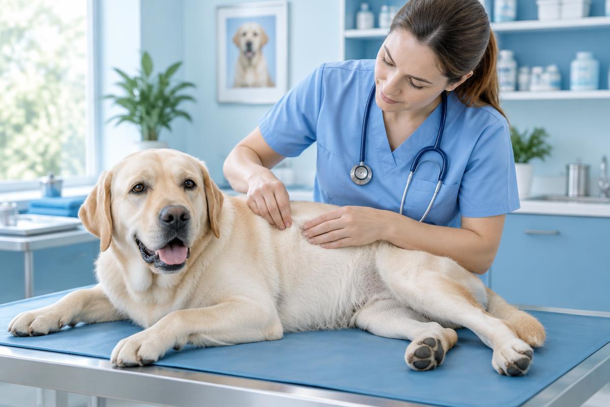
A mast cell tumor, also called a mastocytoma, is a growth that commonly develops in a dog's skin or underlying tissue. Appearance alone cannot establish its identity or risk. Examination and fine-needle aspiration are usually the starting point; carefully planned surgery is often the main treatment for a localized tumor.
Not every mast cell tumor behaves aggressively. Prognosis depends on tumor type, grade, stage and the individual dog. Even a small, apparently harmless growth deserves assessment before an unplanned removal.
Key points
- These tumors can resemble warts, fatty lumps or insect bites.
- Swelling can make their size fluctuate; do not squeeze, rub or massage them.
- Cytology often identifies the tumor; histology assesses grade and surgical margins.
- Lymph nodes may need testing even when they feel normal.
- Treatment and monitoring are individualized, not determined by the diagnosis alone.
What is a mast cell tumor?
Mast cells are normal immune cells involved in inflammation and allergic responses. They contain histamine and other active substances. When these cells become cancerous, they multiply abnormally. Release of their granule contents can cause local swelling and sometimes effects elsewhere in the body.
Cutaneous and subcutaneous tumors
Cutaneous tumors arise within the skin; subcutaneous tumors develop in the tissue beneath it. Touch alone cannot reliably distinguish them. Tissue examination establishes their location. Grading systems designed for cutaneous tumors do not apply directly to subcutaneous tumors, which require assessment of their own risk features.
Boxers, Boston terriers, pugs, Labrador retrievers, golden retrievers and Shar-Pei are among breeds reported to have increased susceptibility. Any dog, including a mixed-breed dog, can be affected. Breed alone does not determine behavior or outcome.
Why is it called “the great imitator”?
A mast cell tumor may be soft or firm, smooth or ulcerated, hairless or covered with hair. Some remain small for a while; others enlarge quickly. Lack of pain does not rule out a tumor. If you have found a lump on your dog, record when you noticed it and arrange an examination.
A growth may become red and swollen after touching, then shrink again. This reaction, known as Darier's sign, reflects release of substances from mast cells. It is not a diagnosis by itself. Do not deliberately provoke it or puncture the lump at home.
Can it cause general illness?
Histamine release can sometimes contribute to gastrointestinal irritation and ulcers. Vomiting, poor appetite, weakness and black, tarry stools are possible. Serious systemic effects are less common than local changes but need prompt attention. These symptoms also occur with other diseases, so an in-person assessment is essential.
How is the diagnosis confirmed?
Cytology: often the first test
Fine-needle aspiration collects cells for microscopic examination. It often identifies a mast cell tumor without major surgery. Occasionally the sample is inconclusive or the cells are unusual, requiring repeat sampling or biopsy. The guide to cytology versus biopsy explains why these tests answer different questions.
Histology: grade and margins
A pathologist examines tissue after removal. Cutaneous tumors may be graded using Patnaik's three-grade system and Kiupel's two-tier system: low grade or high grade. These describe microscopic features, not lump size. Grade differs from stage, which describes how far disease has spread.
The report also evaluates surgical margins and the mitotic count, a measure of dividing cells in a defined tissue area. Clear margins mean no tumor cells were found at the examined specimen edges, not an absolute guarantee against recurrence. KIT mutation testing or Ki-67 assessment may sometimes help refine risk or treatment choices; neither is necessary for every dog.
Checking for spread
Regional lymph nodes draining the tumor area deserve attention even if they are not enlarged. The veterinarian may recommend aspiration or removal for histology. Sometimes identifying the actual draining node requires additional mapping.
The extent of cancer staging depends on examination findings, grade and symptoms. Blood tests, abdominal ultrasound and, when indicated, spleen or liver cytology may be recommended. A normal ultrasound cannot exclude microscopic disease. Conversely, sampling every organ without considering whether results will change care is not automatically useful.
Treatment options
Surgery and local control
For localized disease, surgery with planned lateral and deep margins is often the main treatment. The plan reflects tumor size, location and biological behavior. There is no single margin suitable for every dog. Planning the first operation carefully helps balance tumor control with tissue function.
If margins are incomplete, further excision of the scar or radiation therapy may be considered. Monitoring alone is appropriate in selected situations after risk assessment, not as an automatic response. Incomplete margins do not make recurrence inevitable, but they require discussion.
Systemic and supportive treatment
For high-grade, metastatic or otherwise difficult-to-control disease, the veterinarian may recommend chemotherapy or tyrosine-kinase inhibitors. Decisions depend on tumor characteristics, general health and available options. Monitoring for side effects is part of treatment.
Intratumoral tigilanol tiglate is available in some countries for carefully selected tumors. It is not a universal substitute for surgery: eligibility depends on location and other features, and treatment produces a wound requiring planned aftercare.
Veterinary-prescribed antihistamines and stomach protection may form part of supportive care. Do not choose medicines yourself or adapt human treatment schedules; safety depends on the dog's condition and other prescriptions.
Prognosis and follow-up
Completely removed low-grade tumors often have a favorable outlook. High-grade tumors generally need closer staging and monitoring. Grade, stage, location, margins and the individual animal all matter; no single result tells the whole story.
Follow-up visits assess the scar, skin and lymph nodes, with further tests as needed. At home, gently inspect the skin and photograph changes without squeezing them. Every new lump in a dog previously treated for a mast cell tumor should be checked. It is not necessarily recurrence, but appearance cannot establish safety.
When to seek veterinary care
A rapidly enlarging, ulcerated or bleeding mass needs prompt in-person assessment. Ongoing bleeding, repeated vomiting, black stools, collapse or sudden marked lethargy warrant a veterinary clinic or emergency service now. Do not wait for an online consultation or attempt home treatment.
Questions to ask your veterinarian
- Is cytology diagnostic, or is a biopsy needed before surgery?
- Which lymph node should be tested, and which staging tests will change the plan?
- What surgical margins are feasible here?
- What do the grade, mitotic count and margins mean together?
- Are further tests or treatment needed, and how often should we return?
Frequently asked questions
Can a mast cell tumor shrink on its own?
Swelling can subside, making the lump look smaller. This does not prove that the tumor has disappeared or remove the need for testing.
Is needle aspiration dangerous?
The veterinarian considers local reaction risks and precautions. Fine-needle aspiration is widely used for diagnosis; owners should never puncture a growth themselves.
Can a photograph establish the diagnosis?
No. Photographs can document change but cannot identify the cells, grade the tumor or determine spread.
Does every dog need chemotherapy after surgery?
No. Some localized low-grade tumors can be managed with local treatment and monitoring. Additional treatment depends on the combined findings.
Conclusion
The useful sequence is to identify the cells, plan treatment, obtain histology and agree on follow-up. This avoids decisions based only on appearance while recognizing dogs that need more intensive care.
Book a veterinary oncology consultation to review results and plan next steps. Consultation does not replace an in-person examination or emergency treatment.
Important
This article is educational, not a diagnosis or individual prescription. Your dog needs an individual examination to choose tests and treatment. Online consultation does not replace an in-person examination.
Sources and further reading
- ACVS: Mast cell tumors (opens in a new tab).
- AAHA: 2026 oncology guidelines for dogs and cats (opens in a new tab).
- AAHA: Tumor diagnostics and staging (opens in a new tab).
- Merck Veterinary Manual: Cancer treatment in animals (opens in a new tab).
- Veterinary Cancer Society: Pet owner resources (opens in a new tab).
Updated 24 October 2026
Related articles

Tumors in dogs
Found a lump on your dog? What to do and when to see a vet
Most lumps on dogs are not dangerous, but you can't tell by looking or touching. What to note at home, when to book a vet visit, and which tests show what the lump really is.
5 min read

Treatment & follow-up
A tumor has returned after surgery in your dog or cat — what should you do?
A new lump near a surgical scar does not always mean the tumor has returned, but it needs checking. Learn about possible causes, surgical margins, and the tests that help guide further treatment.
9 min read
Symptoms & diagnostics
Mammary Tumors in Dogs and Cats: Signs, Diagnosis and Treatment
A lump near a nipple cannot be assessed by touch alone. Learn how mammary tumors differ in dogs and cats, which tests may be needed, and how surgery and follow-up care are planned.
8 min read