Symptoms & diagnostics
How cancer is staged in pets: tests and imaging explained
Staging shows how far a tumour has spread. Which tests are used, how stage differs from grade, and how to choose tests sensibly with your vet.
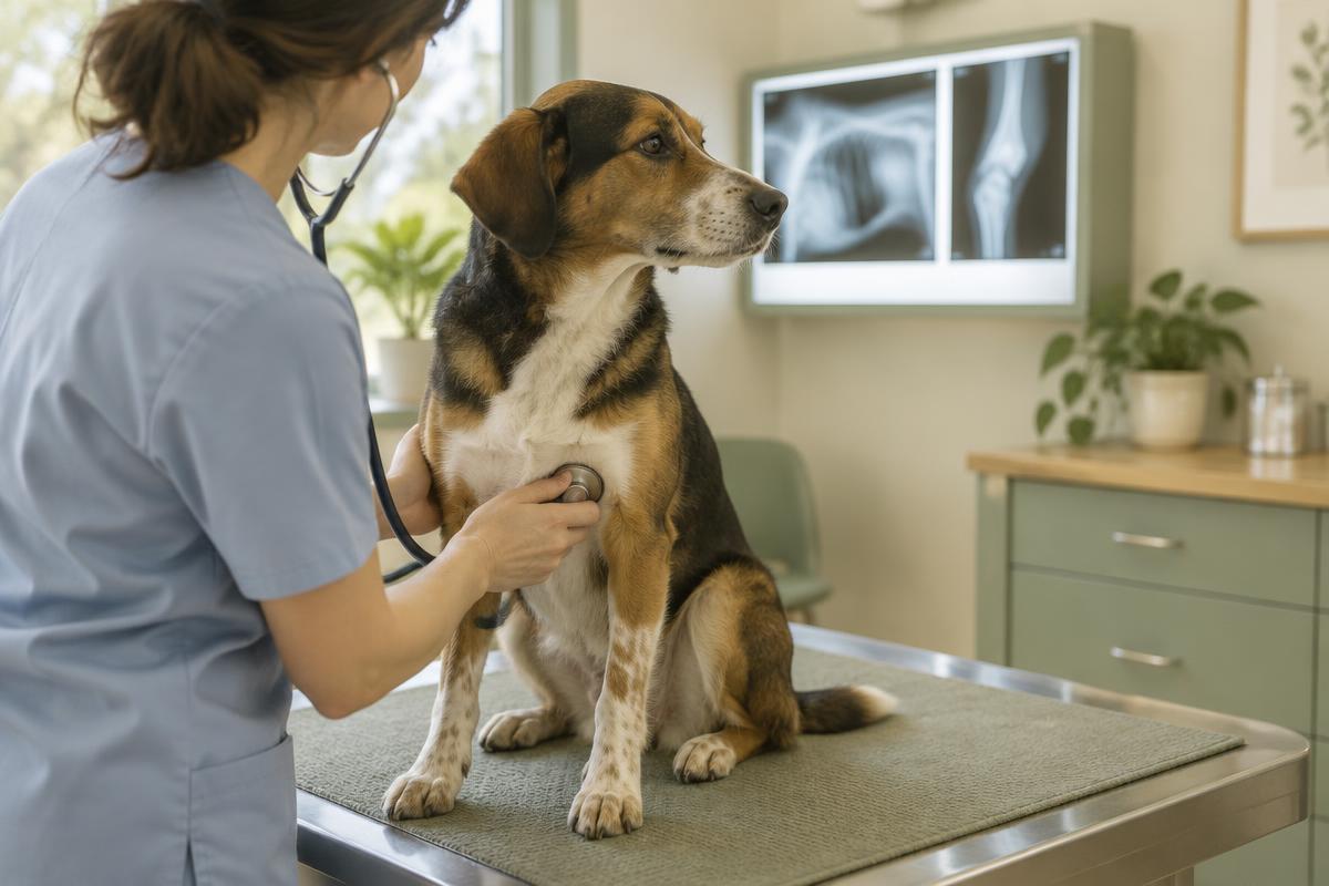
Once a malignant tumour is confirmed, the next question is how far it has spread. The tests that answer it are called staging. Staging helps clarify the outlook and choose treatment — for example, whether surgery alone is likely to be enough or a combined approach is needed. Which tests make sense depends on the tumour type, your pet's overall health and your family's circumstances; not every pet needs every test.
The short answer
- Stage describes the tumour's size and whether it has spread to lymph nodes or other organs.
- Grade describes how the tumour cells look under the microscope. They are different things.
- Staging usually looks at the tumour itself, the nearby lymph nodes, general health and the organs this tumour type tends to spread to.
- A sensible testing plan is agreed together with your vet.
Stage versus grade
These terms are easily confused. Grade is assigned by a pathologist from histopathology: low grade means the cells look fairly normal and invade little, high grade means the cells are very abnormal. Stage comes from the combined test results: a small tumour without spread is early stage; a large, invasive tumour or one that has spread is advanced. Both matter for the outlook. To learn how tissue for histopathology is obtained, see Cytology or biopsy.
What staging involves
- Assessing the tumour itself. The vet measures it and checks whether it moves freely or is fixed to surrounding tissue. If it's internal or may involve bone, ultrasound, X-rays, CT or MRI are used. Ideally, a board-certified veterinary radiologist reviews the images.
- The regional lymph nodes. Feeling them is not enough: an enlarged node may just be reactive, and a normal-sized node can contain cancer cells. That's why a node is often sampled by aspirate or biopsy.
- General health. A minimum database includes a complete blood count, biochemistry and urinalysis, plus testing for feline leukaemia and immunodeficiency viruses in cats. It reveals other conditions that affect treatment and anaesthesia choices.
- Looking for distant spread. Most often chest X-rays in several views or CT, plus abdominal ultrasound. Which organs are checked depends on where that particular tumour usually spreads.
Why the set of tests varies
Oncology guidelines suggest choosing tests by three principles:
- Prognostic — does the test change the outlook or the treatment decision? If not, it may not be needed.
- Practical — if the budget is limited, avoid spending most of it on tests and leaving little for treatment. Prioritise tests that confirm the diagnosis and assess overall health.
- Pertinent — check the sites this tumour spreads to most often. For example, regional lymph nodes are especially important in canine mast cell tumours, and the lungs in osteosarcoma.
Staging systems
Many solid tumours are staged with the TNM system: T for the size and extent of the primary tumour, N for lymph node involvement and M for distant metastasis. Canine lymphoma uses stages I to V with a substage: "a" if the dog feels well, "b" if there are signs of illness. Your vet can explain which system applies and what it means for your pet.
Restaging
Tests are repeated during and after treatment to assess the response and catch any recurrence early. Your vet will advise on the timing of follow-up visits.
Questions to ask your vet
- Which tests are needed for this tumour type, and why?
- How would different results change the treatment plan?
- Does any test require anaesthesia, and how will that risk be assessed?
- Can we prioritise tests if the budget is limited?
- Who will interpret the imaging?
Frequently asked questions
Is staging needed if we choose palliative care?
Not always. Sometimes basic tests are enough to manage symptoms and comfort. Ask your vet which tests would genuinely help in your situation.
Does a benign tumour need staging?
Usually not — benign tumours don't spread. General health is still assessed before surgery.
What if spread is found?
It's important information for choosing treatment, but it isn't always the end of options. What can be done for treatment and quality of life depends on the tumour type and your pet's condition.
Important
This article is for general information and does not replace advice from a veterinarian. A testing plan is always individual. An online consultation with a veterinary oncologist can help you understand results and discuss next steps, but it does not replace an in-person examination.
Sources and further reading
- AAHA Section 3: Tumor Diagnostics and Staging (opens in a new tab)
- AAHA: 2026 Oncology Guidelines for Dogs and Cats (opens in a new tab)
- ACVS: Mast Cell Tumors (opens in a new tab)
- ACVS: Mammary Tumors (opens in a new tab)
- Merck Veterinary Manual: Cancer Treatment in Animals (opens in a new tab)
- Veterinary Cancer Society: Pet Owner Resources (opens in a new tab)
Updated 20 October 2026
Related articles
Symptoms & diagnostics
Mammary Tumors in Dogs and Cats: Signs, Diagnosis and Treatment
A lump near a nipple cannot be assessed by touch alone. Learn how mammary tumors differ in dogs and cats, which tests may be needed, and how surgery and follow-up care are planned.
8 min read

Symptoms & diagnostics
Tumor biopsy in dogs and cats: what happens and why it matters
A biopsy helps identify a tumor and guide treatment. Learn how tissue is collected, what to expect on the day, how to care for the wound, and what the pathology report can tell you.
7 min read

Symptoms & diagnostics
Cancer symptoms in dogs and cats: what to watch for
Signs of cancer in pets are often vague — weight loss, tiredness, a cough or a limp. Which changes to show your vet, which need urgent care, and how to keep useful notes at home.
4 min read