Symptoms & diagnostics
Mammary Tumors in Dogs and Cats: Signs, Diagnosis and Treatment
A lump near a nipple cannot be assessed by touch alone. Learn how mammary tumors differ in dogs and cats, which tests may be needed, and how surgery and follow-up care are planned.
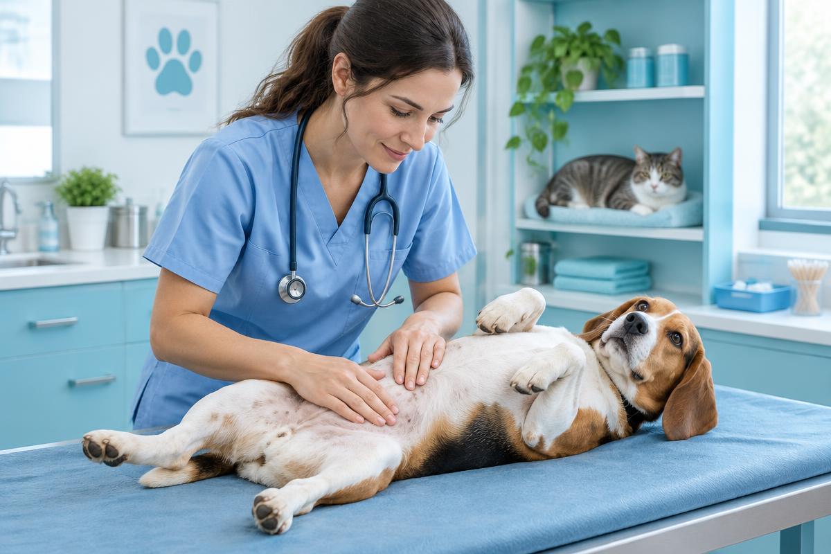
A mammary tumor in a dog or cat warrants an in-person veterinary examination, even if the lump is small and does not bother the animal. Its appearance and firmness cannot reliably reveal what it is. Surgery is the main treatment for operable tumors, and histopathological examination of the tissue provides essential information.
In dogs, approximately half of mammary tumors are benign, although this proportion varies between studies. In cats, most mammary tumors are malignant carcinomas. Any lump along a cat’s mammary chain should therefore be examined promptly, and a growing lump in any animal should not simply be “watched until it gets worse.”
Key points
- Mammary tumors are particularly common in intact females and those spayed later in life.
- Even a small, painless lump needs assessment.
- Cytology is useful but cannot always distinguish a benign mammary tumor from a malignant one.
- The extent of surgery depends on the species, the location of the lumps and the extent of the disease.
- Prognosis depends on the tumor’s type, grade, size and stage, as well as the animal’s health.
- Difficulty breathing or sudden, severe weakness requires immediate veterinary care.
Why mammary tumors develop
Mammary tumors are among the most common neoplasms in intact dogs and cats and those spayed later in life. Sex hormones influence their development, but hormonal stimulation is not the only cause. These tumors are rare in males, but a lump near a nipple in a male dog or cat also needs assessment.
How spaying affects risk
In dogs, the protective effect of spaying is greatest when it is performed before the first heat cycles. However, the precise reduction in risk varies between studies. In cats, early spaying is also associated with a lower risk of mammary tumors.
The timing of surgery is individualized, taking into account the animal’s breed, size and health, as well as other potential consequences of early surgery. This decision should be discussed with a veterinarian rather than based solely on preventing one disease. Spaying will not make an existing tumor disappear.
Changes you may notice
Owners most often find a firm lump near or between the nipples. There may be several lumps, on one or both sides. Swelling, skin changes, ulceration, bleeding and nipple discharge may also occur. A lack of pain does not mean the mass is harmless.
If you find a lump on your dog, note when you first noticed it and any changes in size. Any lump on your cat along the mammary chain needs particularly prompt examination. Do not squeeze out discharge or massage the mass.
Inflammatory mammary carcinoma in dogs
This rare but aggressive form of the disease can resemble mastitis: the mammary area becomes red, hot, painful and diffusely swollen, without a clearly defined lump. These conditions cannot be distinguished from a photograph.
These signs require an urgent in-person examination. If inflammatory mammary carcinoma is confirmed, surgery is usually not the first choice because of the way the disease spreads and the risk of complications. An oncologist will determine the treatment and symptom-relief plan.
How the diagnosis is made
Examination and cell sampling
The veterinarian examines and palpates both mammary chains, measures the masses, and assesses the skin, tissue mobility and lymph nodes. Relevant history includes heat cycles, spaying, hormonal medication use, how quickly the lumps have grown and the animal’s general well-being.
Fine-needle aspiration collects cells for examination. Cytology helps rule out other types of masses and inflammation, and can also be used to assess lymph nodes. However, mammary tumors can be heterogeneous: a small sample may not represent the entire tumor. Cytology therefore cannot reliably distinguish all benign and malignant forms.
Why histopathology is needed
Histopathological examination of the removed tissue is a key test. It identifies the tumor type and grade, evidence of invasion into vessels, and the status of the surgical margins. Sometimes a tumor biopsy is needed before the main surgery if the result would change the treatment approach.
When there are several lumps, it is important to examine them separately: different types of masses may occur in the same animal at the same time. Clean margins mean that no tumor cells were found at the specimen’s edges in the sections examined, but they do not guarantee that the tumor will not recur.
Checking whether the disease has spread
Cancer staging helps establish how far the disease has spread and what extent of treatment is appropriate. Tests commonly discussed include:
- Three-view chest radiographs or computed tomography (CT).
- Assessment of regional lymph nodes, with cytology or histopathology when needed.
- Blood tests to assess general health and prepare for anesthesia.
- Abdominal ultrasound when indicated.
A normal-sized lymph node does not rule out metastasis. Chest imaging also cannot detect every microscopic site of spread. The diagnostic workup is tailored to how the results would affect treatment.
The size of the primary tumor has prognostic significance, especially in cats. This is one reason not to delay seeking care until a small lump interferes with walking or becomes ulcerated.
How mammary tumors are treated
Surgery and its extent
Surgical removal is the main treatment for localized, operable tumors. In dogs, surgery may involve removing an individual lump with appropriate margins, a single gland, a group of glands or the entire mammary chain. The extent of surgery depends not only on size but also on the location and number of masses and whether complete removal is feasible.
In cats, more extensive surgery is often recommended: removal of the entire mammary chain on one side, or removal of both chains in staged procedures. This reflects the way mammary carcinomas spread. Removing only the visible lump often does not provide adequate local control.
Spaying at the time of tumor surgery is considered individually. There may be separate reasons to recommend it, but it does not replace tumor removal or guarantee that the tumor will not return.
Additional treatment
Chemotherapy may be discussed for high-risk malignant tumors, lymph node involvement or metastasis. Its expected benefit depends on the diagnosis and clinical situation; not every animal needs it.
If surgery is not appropriate, the veterinarian will discuss other options for disease control and palliative care. Pain relief, nutritional support and care of damaged tissues are important at every stage. Owners must not choose anticancer medications or human pain medicines for their pets themselves.
Home care after surgery
Before discharge, owners receive a written plan for pain relief, home care and rechecks. An Elizabethan collar or a veterinarian-approved recovery suit, restricted activity and daily wound checks are usually needed.
Do not allow your pet to lick the incision or apply products that have not been prescribed. Increasing redness, swelling, an unpleasant odor, discharge, separation of the wound edges or marked pain requires contact with the clinic and an in-person assessment.
Prognosis and follow-up
Prognosis depends on the tumor’s type and grade, its size, surgical margins, lymph node involvement, distant metastases and the animal’s individual health status. The word “carcinoma” alone is not enough to provide a precise prognosis.
After treatment, follow-up examinations of the mammary glands, surgical scar and lymph nodes are needed; repeat tests are scheduled according to risk and symptoms. At home, it is helpful to gently check the skin and note any new lumps. These do not always mean the original tumor has returned, but each one needs assessment.
When to see a veterinarian
A rapidly growing, ulcerated, bleeding or infected mass should be examined as soon as possible. A red, hot, painful swelling in the mammary area needs an urgent examination that same day. A new cough, refusal to eat or noticeable weakness also warrants prompt in-person assessment.
If your pet has difficulty breathing, collapses, develops sudden severe weakness or has bleeding that will not stop, go immediately to a veterinary clinic or emergency service. Do not wait for an online consultation or try to treat your pet at home.
Questions to ask your veterinarian
- Which masses and lymph nodes need testing?
- Is a biopsy needed before surgery?
- Which tests would change the treatment plan?
- Why is this particular extent of surgery recommended?
- What did the histopathology show, and is additional treatment needed?
- How should pain relief, wound care and follow-up be managed?
Frequently asked questions
Can I wait if the lump is small?
It is best not to delay assessment. Small size does not prove that a lump is benign, and timely evaluation helps with treatment planning.
Could it be mastitis rather than a tumor?
Yes, inflammation and other changes can sometimes look similar. An examination and diagnostic tests help distinguish them, rather than appearance alone.
Does every cat need both mammary chains removed?
No. Extensive surgery is often needed, but its extent and whether it should be staged are determined individually after assessment.
Should an older animal have surgery?
Age alone does not rule out surgery. Concurrent illnesses, anesthetic risk, the extent of the tumor and expected quality of life are more important considerations.
Conclusion
For a mammary lump, a useful sequence is an examination, staging tests, a well-founded treatment plan and histopathology. There is no need to wait for pain or ulceration: seeking care promptly helps preserve more options for treatment and support.
Book a consultation with a veterinary oncologist to help understand the results and plan the next steps. A consultation does not replace an in-person examination or emergency care.
Important information
This material is for educational purposes and does not provide a diagnosis or an individualized treatment recommendation. A veterinarian determines the appropriate tests, treatment and follow-up schedule after assessing the animal.
Sources and further reading
- ACVS: Mammary tumors (in English) (opens in a new tab).
- AAHA: 2026 Oncology Guidelines for Dogs and Cats (in English) (opens in a new tab).
- AAHA: Tumor diagnostics and staging (in English) (opens in a new tab).
- Merck Veterinary Manual: Cancer treatment in animals (in English) (opens in a new tab).
- Veterinary Cancer Society: Resources for pet owners (in English) (opens in a new tab).
Updated 24 October 2026
Related articles
Symptoms & diagnostics
Tumor biopsy in dogs and cats: what happens and why it matters
A biopsy helps identify a tumor and guide treatment. Learn how tissue is collected, what to expect on the day, how to care for the wound, and what the pathology report can tell you.
7 min read
Symptoms & diagnostics
Cancer symptoms in dogs and cats: what to watch for
Signs of cancer in pets are often vague — weight loss, tiredness, a cough or a limp. Which changes to show your vet, which need urgent care, and how to keep useful notes at home.
4 min read
Symptoms & diagnostics
Cytology or biopsy: how tumour tests differ
Cytology and histopathology answer different questions. What each test shows, how long results take, why a biopsy before surgery can matter, and what to do if the answer is unclear.
5 min read