Treatment & follow-up
A tumor has returned after surgery in your dog or cat — what should you do?
A new lump near a surgical scar does not always mean the tumor has returned, but it needs checking. Learn about possible causes, surgical margins, and the tests that help guide further treatment.
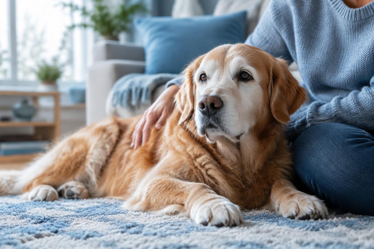
If your dog or cat develops a new lump near the scar after tumor removal, arrange an in-person veterinary examination without delay. This does not necessarily mean recurrence: possible causes include fluid accumulation, a reaction to suture material, scar tissue, or infection. However, these cannot be distinguished from a tumor by a photograph or touch alone.
Do not wait for the lump to grow or become painful. Having it checked does not necessarily mean your pet will need another operation. First, it is important to understand what has changed and compare the findings with the original histopathology report.
Key points
- A new lump near a surgical scar always needs checking, even if it is small and painless.
- Local recurrence, lymph node involvement, and distant metastases are different situations.
- Keep the original histopathology report and the report from the first operation.
- “Clear margins” are reassuring but do not guarantee that a tumor will not recur.
- Further treatment is chosen based on examination and test results, taking your pet’s well-being and quality of life into account.
- Difficulty breathing, collapse, or severe bleeding requires emergency care now.
What can develop near a surgical scar?
Not every lump is a tumor
A seroma—a collection of tissue fluid—sometimes develops after surgery. Other possibilities include firm scar tissue, an inflammatory reaction to sutures, or a granuloma, a nodule that forms around suture material. Redness, pain, and discharge may accompany an infection. Sometimes a different tumor, unrelated to the original one, develops nearby.
The timing helps the veterinarian assess possible causes, but it does not establish a diagnosis. Even a long-healed scar can change for reasons unrelated to cancer, while a small, painless lump may be a tumor.
Recurrence and spread are not the same thing
Local recurrence means regrowth of the original tumor at the surgical site. Regional spread means involvement of nearby lymph nodes or those that drain the affected area. Distant metastases are tumor deposits in distant organs or tissues.
A new, unrelated tumor develops independently of the original disease. Distinguishing between these situations requires testing and sometimes comparison of the old and new tissue samples. A lump near a scar does not, by itself, prove that cancer has spread throughout the body.
Why a tumor may return
Microscopic cells beyond the visible boundary
Some tumors extend between normal tissues farther than the surgeon can see or feel. This can occur with soft tissue sarcomas, mast cell tumors, and feline injection-site sarcomas. The behavior of an individual tumor depends on its type and grade.
Microscopic tumor cells may remain after removal. If they resume growing, the tumor can recur. This is possible even when the scar looks completely normal immediately after surgery.
Limitations of the first operation
The risk may be higher after incomplete removal or when surgical margins are narrow. Sometimes the tumor type is not known before surgery, so the necessary extent of surgery is not planned in advance. In other cases, important blood vessels, nerves, or organs nearby limit how much surrounding tissue can be removed.
Recurrence alone does not prove that the surgeon made a mistake. Understanding what happened requires the original records and information about the tumor’s location and biological behavior.
Understanding surgical margins
What the pathologist examines
Surgical margins are the outer boundaries of the removed tissue specimen. The pathologist checks for tumor cells near these boundaries and measures the distance between the tumor and the margin.
- Clear, or tumor-free, margins: no tumor cells were found at the edge of the specimen in the sections examined.
- Close, or narrow, margins: tumor cells are near the edge; the margin width stated in the report matters.
- Incomplete margins: tumor cells extend to the edge of the specimen, raising concern that tumor tissue may remain in the body.
There is no single safe margin width for every tumor. Both the lateral and deep margins matter, as do the types of tissue between the tumor and the boundary and the characteristics of the tumor itself.
Why the original report matters
The phrase “it was all removed” is not a substitute for a histopathology report. The report may specify the exact tumor type, grade, cell division activity, and distance to the margins.
Clear margins do not provide an absolute guarantee: selected tissue sections are examined, not every cell in the specimen. Incomplete margins do not mean recurrence is inevitable, either, but they do require discussion of the next steps. If needed, the oncologist may recommend having another pathologist review the slides.
What owners should do, step by step
- Do not squeeze or puncture the lump. Do not massage the scar, apply heat, or use any products unless prescribed.
- Take a photograph with a ruler beside the lump. Record the date, approximate size, and any changes in the skin or your pet’s well-being. Do not press the ruler against the lump.
- Gather the records. These include histopathology and cytology reports, the surgical report, discharge summaries, diagnostic images and reports, blood test results, and a list of prescribed treatments.
- Contact the clinic and arrange the earliest available in-person examination. Explain that the lump appeared after cancer surgery and describe any symptoms.
Photographs are useful for comparison, but they do not replace diagnostic testing. Do not delay the visit to collect more photographs or find every document: missing records can be requested later.
Which tests may be needed?
Examination and testing of the lump
The veterinarian will assess the scar, the lump’s mobility, the skin, any tenderness, and the lymph nodes. Fine-needle aspiration with cytology is often the first test. It helps identify the types of cells present but does not always provide a definitive answer.
If there is too little material, or if the tissue architecture needs to be assessed, a biopsy with histopathology is needed. Read more about why these tests are not interchangeable in Cytology and biopsy. Ideally, the biopsy site should be agreed with the surgeon beforehand so that it does not complicate later removal.
Restaging
When recurrence is confirmed or there is a well-founded suspicion, the veterinarian reassesses the extent of the disease. The plan may include lymph node testing, chest imaging, abdominal ultrasound, and laboratory tests to assess overall health.
Sometimes CT helps determine the depth of involvement and plan surgery. Not every animal needs the same set of tests: cancer staging should answer questions that genuinely affect treatment decisions. A normal-sized lymph node does not rule out tumor involvement, and normal imaging does not rule out microscopic disease.
Which treatment options may be discussed?
Repeat surgery and local treatment
If the disease is localized and surgery is appropriate, a surgical oncologist may plan a wider excision of the lump, scar, and surrounding tissues. Simply removing the visible nodule again does not always provide adequate local control.
Radiation therapy may be considered for local control, particularly after incomplete removal when further surgery is not possible or would be too invasive. Electrochemotherapy is available in selected cases. It is not suitable for every tumor and requires specialized equipment and an assessment of whether it is appropriate.
Systemic therapy and supportive care
For certain tumor types, a high risk of spread, or metastatic disease, chemotherapy and other systemic treatments may be discussed. These are not prescribed automatically simply because a tumor has “recurred,” and they do not always replace local treatment.
Palliative care focuses on pain relief, wound care, and maintaining nutrition and comfort. It is active medical care, not giving up on your pet. Every option takes into account quality of life, the expected benefit, the burden of treatment, and the family’s resources and circumstances.
Prognosis depends on the tumor type, grade, stage, and the individual animal’s condition. A second opinion can be helpful before complex repeat surgery or when results conflict; it should not delay urgent care.
When to seek urgent care
A rapidly growing, ulcerated, or bleeding lump needs a prompt in-person examination. If the wound opens, your pet develops a fever or a new cough, or stops eating, contact the clinic the same day.
Difficulty breathing, collapse, sudden severe weakness, or ongoing significant bleeding means you should go to the nearest 24-hour veterinary clinic now. Do not wait for an online consultation or try to treat these conditions at home.
Follow-up after treatment
The follow-up schedule is tailored to the individual animal. Checks are usually more frequent early on and when the risk is high. At these visits, the veterinarian examines the scar, skin, and lymph nodes; imaging is repeated when indicated.
At home, a gentle check without squeezing the tissues is enough. Any new lump is a reason to contact your veterinarian, even if the next scheduled checkup is some time away. Ask for a written follow-up plan with dates and a list of symptoms that should not wait.
Questions to ask your veterinarian
- Has recurrence been confirmed, or could there be another cause?
- What does the original histopathology report say about the margins and their width?
- Are a slide review and restaging needed?
- What is the goal of each treatment option, and what burden does it involve?
- How will we assess comfort and treatment results?
Frequently asked questions
Can I wait if the lump is small?
No. Size and the absence of pain cannot tell you what the lump is. Arrange an examination rather than waiting for it to grow.
Do clear margins rule out the tumor returning?
No. They are an important favorable finding, but they must be considered alongside the tumor’s type, grade, and stage.
Is another operation always necessary?
Not always. The decision depends on the diagnosis, the extent of the disease, whether removal is feasible, and your pet’s condition.
Conclusion
A new lump near a surgical scar is a reason for investigation, not a diagnosis in itself. The original histopathology report, testing of the lump, and a carefully considered plan help guide the next steps without rushed decisions.
You can book a veterinary oncology consultation to better understand the results and plan the next steps. An online consultation does not replace an in-person examination or emergency care.
Important information
This article is for educational purposes and does not provide an individual diagnosis or treatment prescription. A veterinarian selects diagnostic tests and treatment after assessing the animal. Do not start or change treatment on your own.
Sources and further reading
- AAHA: 2026 Oncology Guidelines for Dogs and Cats (in English) (opens in a new tab).
- AAHA: Tumor Diagnostics and Staging (in English) (opens in a new tab).
- AAHA: Supportive and Symptomatic Care (in English) (opens in a new tab).
- Merck Veterinary Manual: Cancer Treatment in Animals (in English) (opens in a new tab).
- Veterinary Cancer Society: Pet Owner Resources (in English) (opens in a new tab).
Updated 24 October 2026
Related articles
Treatment & follow-up
Chemotherapy for dogs and cats: treatment and side effects
Veterinary chemotherapy aims to control cancer while preserving a good quality of life. Learn what happens at appointments, which side effects to watch for, and when your pet needs urgent care.
7 min read
Symptoms & diagnostics
Mammary Tumors in Dogs and Cats: Signs, Diagnosis and Treatment
A lump near a nipple cannot be assessed by touch alone. Learn how mammary tumors differ in dogs and cats, which tests may be needed, and how surgery and follow-up care are planned.
8 min read
Symptoms & diagnostics
Tumor biopsy in dogs and cats: what happens and why it matters
A biopsy helps identify a tumor and guide treatment. Learn how tissue is collected, what to expect on the day, how to care for the wound, and what the pathology report can tell you.
7 min read